
Facets
Browse across eight MeSH (opens in new tab) facets — era, geography, science, specialty, technology, history, culture, and reference. Select one tag per group; counts update across the others. What’s new in facet browse how facets relate to subjects and MeSH.
Clear filtersFacet filters
Geography
Specialties & Disease
- Anatomy & Pathology 199
- Cardiology & Blood 9
- Neurology & Psychiatry 55
- Obstetrics & Reproductive 30
- Infectious Disease (General) 0
- Surgery & Anesthesia 22
- Public Health 30
- Immunology & Dermatology 19
- General Clinical Medicine 25
- Military Medicine 3
- Psychology 14
- Alternative & Fringe Medicine 27
- Pediatrics 5
- Ophthalmology & Vision 5
- ENT & Hearing 7
- Urology & Nephrology 7
- Gastroenterology & Hepatology 1
- Pulmonary & Respiratory 3
- Rheumatology, Rehab & Pain 2
- Internal, Emergency & Geriatric 0
- Veterinary Medicine 6
- Epidemiology & Demography 18
- Physiology & Embryology 17
- Dentistry 10
- Plagues & Epidemics 27
- Microbiology & Virology 1
Social & Historical Studies
Institutions & Culture
Reference & Scholarly Works
Drugs & Technology
199 entries match Anatomy & Pathology [G02.149 / C23] · Arts, Literature & Humanities [K01.090]
1741
#395.2
Tabulae anatomicae.
![PIETRO DA CORTONA, [Pietro BERRETINI] (1596 – 1669)](/media/portraits/743)
27 anatomical copperplates after drawings by the most influential painter of the Italian Baroque movement, who also excelled as an architect. The editor, Cajetano Petrioli, supplied the text and small numbered anatomi…
1743 –1756
#397
Icones anatomicae. 8 pts.
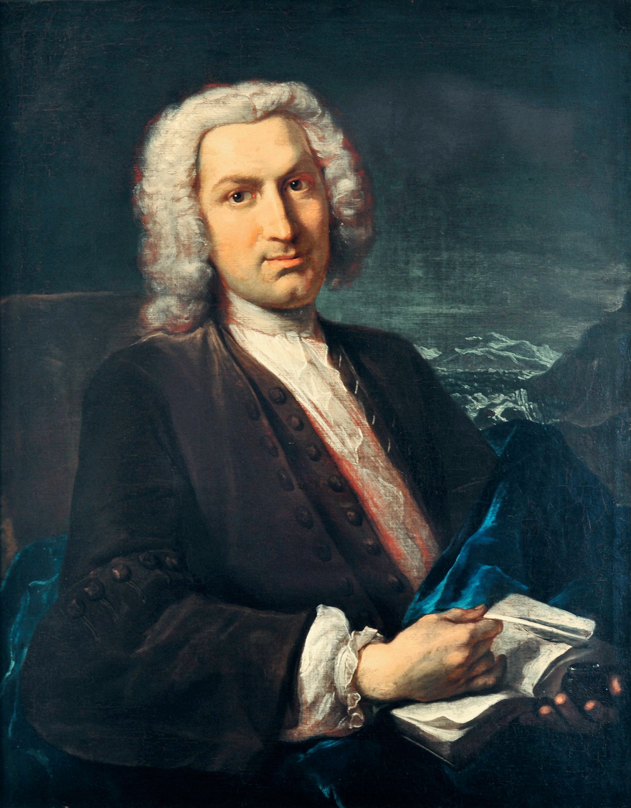
Accurate and beautiful engravings of the diaphragm, uterus, ovaries, vagina, arteries, with explanatory observations. About fifty years after they were originally published the most visually spectacular versions of Ha…
1745
#398
Essai d’anatomie en tableaux imprimés, qui represent au naturel tous les muscles de la face, du col, de la tête, de la langue & du larinx. d'après les parties disséquées & préparées par Monsieur Duverney....comprenant hit grandes planches.
Remarkable for its striking mezzotints printed in color. Digital facsimile from the Internet Archive at this link.
1748
#7491
Anatomie de la tête, en tableaux imprimés qui représentent au naturel le cerveau sous différentes coupes, la distribution des vaisseaux dans toutes les parties de la tête, les organes des sens et une partie de la névrologie, d'après les pièces disséquées et préparées par M. Duverney, en huit grandes planches dessinées, peintes, gravées et imprimées en couleur et grandeur naturelle....
Includes eight spectacular plates printed in color by Gautier d'Agoty with text by Duvereny. Digital facsimile from the Internet Archive at this link.
1754
#7492
Anatomie generale des visceres, et de la nevrologie, angeologie et osteologie du corps humain, en figures, de couleurs et grandeurs naturelles dediée et présentée.
Includes 18 full-page color-printed mezzotints, 12 of the plates designed to fit together in threes to make four life-size human figures. Gautier credited Mertrud, the King's Surgeon with some of the anatomical work i…
1756
#7489
An essay on the demonstration of the human structure, half as large as nature, in four tables, from the pictures painted after dissections, for that purpose.
Four large mezzotint plates issued with an accompanying text.
1759
#7006
Traité d'ostéologie. 2 vols.
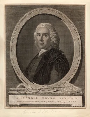
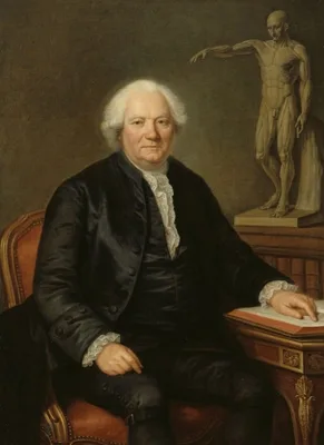
Monro Primus' textbook on the anatomy of the bones was originally published in 1726 as an octavo volume without plates, and went through more than ten editions. The French translation, published in large folio, transl…
1766
#308.1
The anatomy of the horse.
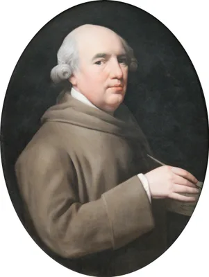
The first original work on equine anatomy after Ruini (No. 285). Stubbs, the great painter of animals, prepared his own dissections of horse carcasses, and personally engraved the 24 double folio plates for this work,…
1771
#6604.9
Verzeichnis einer Samlung von Bildnissen, gröstentheils berühmter Aerzte; so wohl in Kupferstichen, schwarzer Kunst und Holzschnitten, als auch in einigen Handzeichungen: diesem sin verschiedene Nachrichten und Unmerkungen Vorgesekt, die so wohl zur Geschichte der Arzenengelahrtheit; als vornehinsich zur Geschichte der Künste gehören.
“Very valuable reports on artistic anatomy and the history of anatomic illustration” (Choulant). Probably represents the first history of medical portraiture. Digital facsimile from Googe Books at this link.
1773
#155
Théorie de la figure humaine.
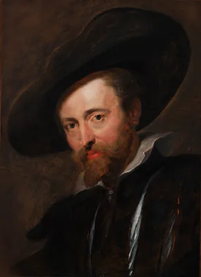
This work on the human figure, published more than 100 years after the death of Rubens, is one of a handful of anatomical treatises illustrated by an artist of the first magnitude.
1775
#399.1
Septemdecim tabulae…
Santorini died before the completion of these anatomical plates which he intended to be his chef d’oeuvre. This elegantly printed volume is the only significant medical book printed by the celebrated Giambattista Bodo…
1777 –1781
#2278
Observationes anatomicae-pathologicae. 4 vols.
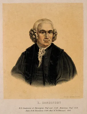
Sandifort’s beautifully illustrated work on pathological anatomy included records of ulcerative aortic endocarditis, renal calculi, hemias, bony ankyloses, and congenital abnormalities. A good account of the “tetralog…
1778
#1383
Dissertatio inauguralis anatomica de basi encephali et originibus nervorum cranio egredientium libri quinque.
The first accurate enumeration of the 12 cranial nerves, superseding that of Willis (No. 1378). Soemmerring is notable for his accuracy in anatomical illustration. This was his thesis. The same publisher issued an edi…
1779
#401.1
Nouveau recueil d’ostéologie et de myologie…
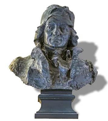
“Without contest the most beautiful of all anatomies for the artist and one of the most remarkable books of its time” (Hahn & Dumaitre). The plates in this work are more fantastic than any other anatomy, suggesting th…
1780
#9547
Osteografia e miografia della testa, mani, e piedi del corpo umano in misura naturale con il catalogo de' nomi propri.
Very distinctive engravings with the captions included within the engraved surfaces. Printed and sold by the artist.
1787
#1104
Vasorum lymphaticorum corporis humani historia et ichnographia.
Mascagni, Professor of Anatomy at Siena, made several discoveries regarding the lymphatics. His beautiful atlas contained 41 engravings of the lymphatics and gained him lasting fame. He had previously published a Prod…
1787
#12163
A collection of engravings, tending to illustrate the generation and parturition of animals, and of the human species.
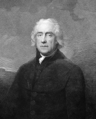
An idiosyncratic collection of rarely reproduced images with explanatory commentaries in English and French, concerning reproduction and obstetric complications in animals and humans. Topics include: The Funis of a nu…
1793 –1835
#11312
Museum anatomicum academiae Lugduno-Batavae. 4 vols.
This work, complete in over 1000 pages, with hundreds of full page plates, was begun by Eduard Sandifort and completed 42 years later by his son Gerard. Many of the plates illustrate diseases of bone. Digital facsimil…
1794
#9833
Engravings, explaining the anatomy of the bones, muscles and joints.
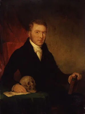
Bell’s atlas of the bones, muscles and joints was issued as a separate work a year after his text, The Anatomy of the Bones, Muscles, and Joints. Bell’s illustrations are some of the most striking in the entire litera…
1797
#401
Tabula sceleti feminini juncta descriptione.
Soemmerring was noted for his accuracy in anatomical illustration, and the above work is a fine example of his artistic sense. For it he selected the skeleton of a well-built girl of 20 years. Great care was taken in …
1797 –1804
#401.3
The anatomy of the human body. 4 vols.
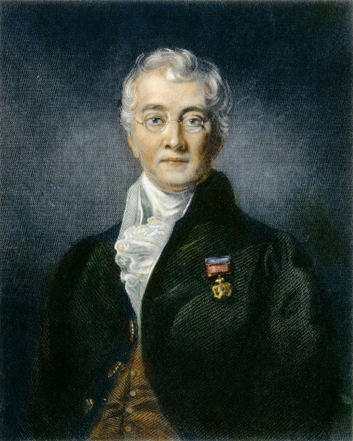
“The first great textbook contributed by the British school to modern anatomy” (Russell, No. 461).
1798 –1803
#402
A system of dissections. 2 vols.
Published in 7 fascicules and appendix while Bell was still a student, this was Bell’s first independent venture as an author. The anatomical work of Charles Bell and his brother John was among the most significant in…
1799 –1803
#2282
A series of engravings, accompanied with explanations, which are intended to illustrate the morbid anatomy of some of the most important parts of the human body.
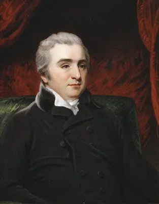
The first systematic atlas of pathology. This work was intended to illustrate No. 2281, but, with its extensive descriptive text for each plate, it may be appreciated separately. The black & white engravings were prep…
1801
#1489
Abbildungen des menschlichen Auges.
Soemmerring is best remembered for his fine anatomical illustrations, of which those devoted to the human eye are a good example. In 1791 he made important observations on the macula lutea: "De foramine centrali limbo…
1806
#1554
Abbildungen des menschlichen Hoerorganes
1806
#6604.92
Essays on the anatomy of expression in painting.
Bell’s artistic and literary skills combined with his knowledge of anatomy and physiology to make this work a tour de force of art history and the anatomical and physiological basis of facial expression.
1811
#406
Anatomia per uso de’pittori e scultori.
This anatomy for artists and sculptors contains 38 good copperplates in black and red.
1812
#14179
Anatomie du gladiateur combattant, applicable aux beaux arts . . . .
Salvage's 21 plates after his own drawings "are based on three casts of bodies dissected to different anatomical layers and set in the pose of the Borghese Gladiator. For these casts he preferred to use the bodies of …
1813
#13214
Kaitai hatsumo [Explanation of Human Anatomy]. 5 vols.
The first Japanese exposition of the whole system of human anatomy, based on original observation. The work includes the records of three dissections performed in Kyoto in 1783, 1798, and 1802. “A Japanese treatise on…
1816
#7488
Anatomia per uso degli studiosi di scultura e pittura.
Mascagni's anatomy for artists and sculptors, edited for posthumous publication by Mascagni's literary execultor Francesco Antonmarchi. 15 hand-colored engraved plates after drawings by Antonio Serantoni (1780j-1837),…
1817
#12746
Nosologie naturelle, ou les maladies du corps humain distribuées par familles. Tome premier. (All published.)
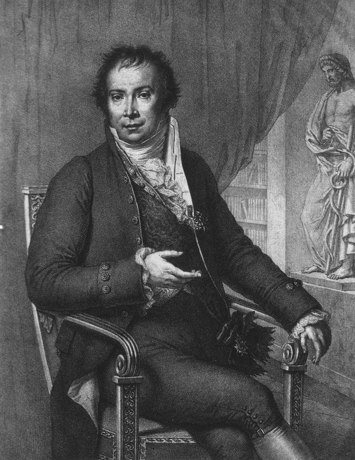
This unfinished work with 22 spectacular plates included some full body images of patients, and unlike other illustrated works by Alibert, it concerned more than skin diseases. Vol. 2 was never published but a second,…
1818
#408
Afbeeldingen van de juiste plaatsing der inwendige deelen van het menschelijk ligchaam.
First anatomical illustrations of frozen sections. De Riemer appears to have been the first to freeze tissues in order to permit fine sectioning for the purposes of diagnosing diseased tissue. Digital facsimile from U…
1821 –1831
#409
Anatomie de l’homme, ou descriptions et figures lithographiées de toutes les parties du corps humain. 5 vols.
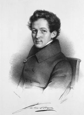
The first anatomical atlas illustrated by lithography, containing 300 plates in folio format. This was one of the most elaborate of the lithographic “incunabula” produced by Charles Philibert de Lasteyrie, one of the …
1822 –1832
#409.1
Anatomia universa… 2 vols.
The largest of all medical books from the standpoint of format. The 44 life size engraved plates are reproduced in double elephant folio size measuring 950 x 635 mm., and include an almost incredible level of detail. …
1822
#13314
System of surgical anatomy. Part first [all published]: On the structure of the groin, pelvis, and perineum. As connected with inguinal and femoral hernia; Tyeing the iliac arteries; and the operation of lithotomy. Illustrated by nine copper-plate engravings
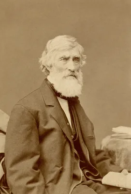
Anderson trained at the Royal College of Surgeons in Edinburgh, emigrated to America in 1820 and, after delivering a series of anatomical lectures in New York under the patronage of Valentine Mott, became affiliated w…
1822 –1826
#13508
A system of anatomical plates of the human body. By John Lizars. Text in 8vo in 12 pts., plus folio atlas with engraved title and 101 plates engraved by William Home Lizars.
Lizars's System of anatomical plates was by far his most successful work, going through many editions. The text of the work was originally issued in 12 parts in octavo format, which were then bound together in book fo…
1823 –1826
#7242
Planches anatomiques du corps humain executes d’après les dimensions naturelles. Double elephant folio atlas and small folio text.
Considering that it is among the rarest of all anatomies, and certainly the largest and probably the most spectacular, it is remarkable that two nearly identical editions of Mascagni’s posthumous life-size anatomy wer…
1826
#2284.1
The morbid anatomy of the human brain.
Based on over 4000 autopsies performed over 30 years, and illustrated with fine hand-colored plates.
1827 –1831
#2285
Reports of medical cases, selected with a view of illustrating the symptoms and cure of diseases by a reference to morbid anatomy. 2 vols. in 3.
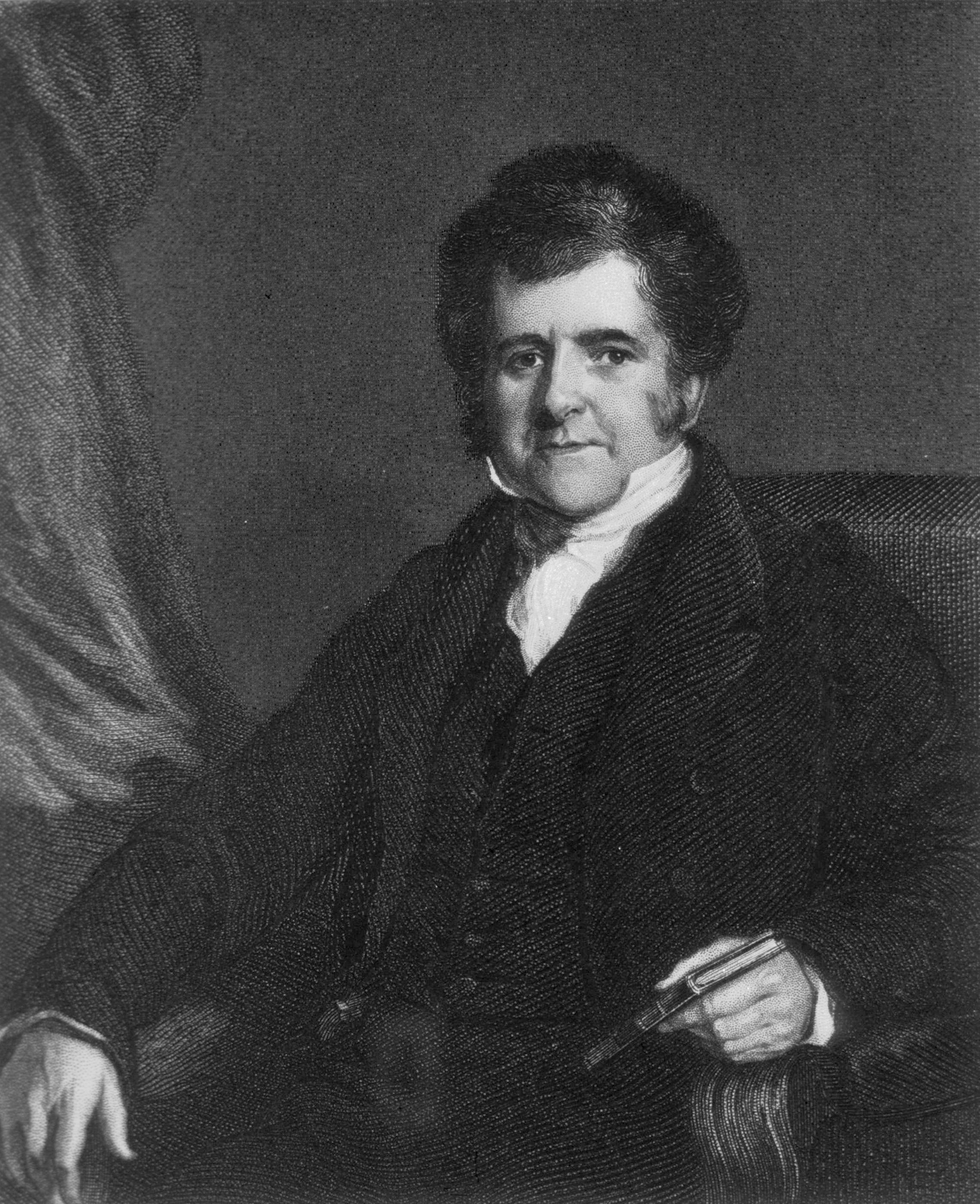
Beside's Bright's classic description of chronic non-suppurative nephritis, known eponymically as “Bright’s disease”, the Reports contain numerous other outstanding contributions to general pathology, neuropathology, …
1828
#2285.1
Traité des maladies des enfans nouveau-nés et à la mamelle. 1 vol. and atlas.
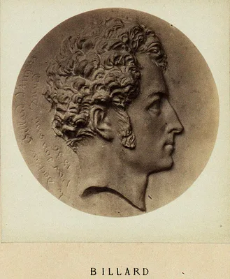
The first significant work on the pathological anatomy of infants. Billard performed several hundred autopsies on infants and children and correlated the data obtained with clinical observations he had made. This pion…
1828
#1776.1
Researches into the causes, nature and treatment of the more prevalent diseases of India, and of warm climates generally. Illustrated with cases, post mortem examinations, and numerous coloured engravings of morbid structures. 2 vols.
A landmark in geographical pathology, superbly illustrated. Annesley’s cases, collected over many years’ service throughout India, represented the most complete treatment of diseases on the sub-continent to date. Digi…
1828
#10023
Myology, illustrated by plates.
This large folio work contains 8 hand-colored lithographed plates by F. R. Say, each with multiple lift-up flaps. It is probably the largest format anatomical work ever published in English with lift-up flaps, with up…
1829 –1842
#2286
Anatomie pathologique du corps humain. 2 vols.
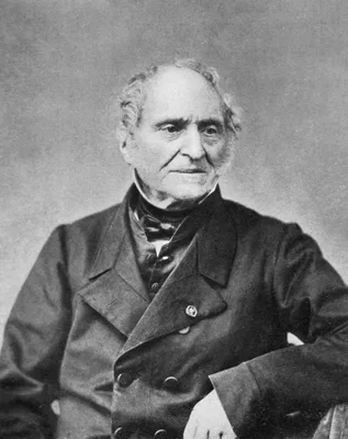
The fine hand-colored lithographs of gross pathology make this one of the greatest works of its kind. Cruveilhier, first Professor of Pathological Anatomy in Paris, gave the first description of multiple sclerosis (in…
1829 –1833
#2288
Traité d’anatomie pathologique. 2 vols. and atlas.
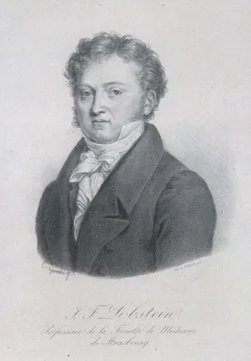
Includes a historical review of the subject from the time of the Ancient Egyptians to Corvisart, and a summary of the advances in pathology during the preceding 50 years. Vol. 2, pp. 553-600 deals with diseases of the…
1829
#11162
Anatomie des formes extérieures du corps humain appliquée à la peinture, à la sculpture et à la chirurgie.
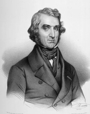
Digital facsimile from the Internet Archive at this link.
1830
#2611
A short tract on the formation of tumours, and the peculiarities that are met with in the structure of those that have become cancerous; with their mode of treatment.
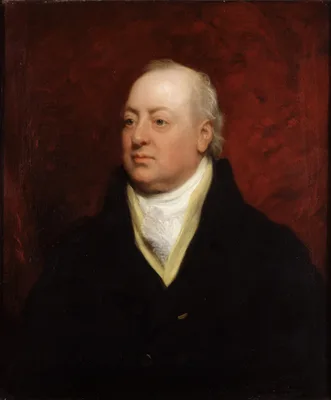
Contains the first illustrations of microscopic sections of cancer; however, Home drew no worthwhile conclusion from his microscopic studies. Digital facsimile from the Internet Archive at this link.
1831 –1854
#7246
Traité complet de l'anatomie de l'homme, comprenant la médecine opératoire. 16 vols.
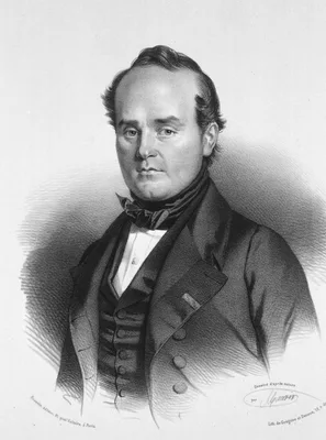
With over 2000 pages of text and 726 lithographed plates (incorporating 3604 individual figures), this work is the most comprehensive, and perhaps the most beautiful anatomical surgical atlas of the 19th century. It w…
1832 –1862
#14348
Atlas der pathologischen Anatomie für praktische Aerzte. 4 parts in 1. With: Erläuterung zu dem Atlasse der pathologischen Anatomie für praktischen Aertze. 4 vols. in 5.
Albers' atlas of pathology, with 257 lithographed plates (93 hand-colored) covers diseases of the brain and spinal cord (Part 1), the throat (Part 2), the thorax (Part 3) and the abdomen (part 4); the five text volume…
1833
#411
Anatomical studies of the bones and muscles, for the use of artists. From drawings by the late John Flaxman, Esq. R.A. Engraved by Henry Landseer. With two additional plates, and explanatory notes, by William Robertson.
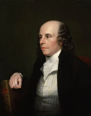
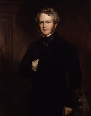
Digital facsimile from digitalcollections.nypl.org at this link.
1833
#411.1
The hand: Its mechanism and vital endowments as evincing design.
Classic work on the anatomy, physiology, bio-mechanics, comparative anatomy, and adaptive importance of the hand. Issued as a volume in a series entitled the "Bridgewater Treatises." The first edition has 288pp. An en…