
Facets
Browse across eight MeSH (opens in new tab) facets — era, geography, science, specialty, technology, history, culture, and reference. Select one tag per group; counts update across the others. What’s new in facet browse how facets relate to subjects and MeSH.
Clear filtersFacet filters
Geography
Specialties & Disease
- Anatomy & Pathology 290
- Cardiology & Blood 3
- Neurology & Psychiatry 19
- Obstetrics & Reproductive 3
- Infectious Disease (General) 0
- Surgery & Anesthesia 14
- Public Health 6
- Immunology & Dermatology 11
- General Clinical Medicine 4
- Military Medicine 0
- Psychology 1
- Alternative & Fringe Medicine 0
- Pediatrics 2
- Ophthalmology & Vision 4
- ENT & Hearing 5
- Urology & Nephrology 12
- Gastroenterology & Hepatology 20
- Pulmonary & Respiratory 1
- Rheumatology, Rehab & Pain 0
- Internal, Emergency & Geriatric 0
- Veterinary Medicine 1
- Epidemiology & Demography 0
- Physiology & Embryology 29
- Dentistry 4
- Plagues & Epidemics 0
- Microbiology & Virology 3
Social & Historical Studies
Institutions & Culture
Reference & Scholarly Works
Drugs & Technology
290 entries match Modern [K01.400.504]
1836
#7674
The anatomist's instructor, and museum companion; being practical directions for the formation and subsequent management of anatomical museums.
"Dr Frederick Knox was the librarian of New Zealand's first public library. He emigrated from Scotland in July 1840 and within days of arriving in Port Nicholson became involved in establishing the country's first pub…
1836 –1842
#13299
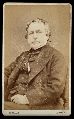
A series of anatomical plates. 5 vols.: The muscles of the human body. The vessels of the human body. The nerves of the human body. The viscera of the human body. The bones and ligaments of the human body.
1836 –1842
#13300
A series of anatomical plates. 5 vols.: The muscles of the human body. The vessels of the human body. The nerves of the human body. The viscera of the human body. The bones and ligaments of the human body.
The most ambitious 19th century English anatomy illustrated by lithography. Some copies were issued with hand-colored plates. The five volumes, containing a total of 201 plates, describe the muscles, blood vessels, ne…
1837 –1841
#11697
Anatomia chirurgica truncorum arteriarum nec non fasciarum fibrosarum.
An atlas of arterial stems and fasciae. Translated into German as Chirurgische anatomie der Arterienstamme und Fascien neu Bearbeitet von Julius Szymanowski. Leipzig und Heidelberg: C. F. Winter, 1850. Digital facsimi…
1838
#7672
Histoire des embaumements et de la préparation des pièces d'anatomie normale, d'anatomie pathologique et de l'histoire naturelle suivie de procédés nouveau.
Gannal discovered the efficacy of solutions of aluminum acetate and chloride for preserving anatomical preparations. His method of embalming involved injection of solutions of aluminum salts into the arteries. Transla…
1840
#542
On the minute structure and movements of voluntary muscle.
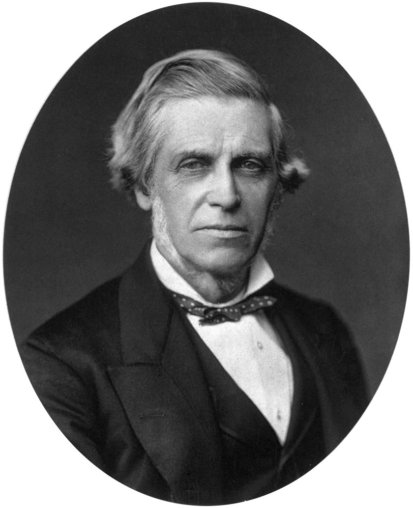
Classical description of striated muscle
1840
#1263
Nuovi organi scoperti nel corpo humano.
“Pacini’s corpuscles”, end organs of sensory nerves, earlier described by Vater in 1717.
1841
#543
Allgemeine Anatomie. Lehre von den Mischungs- und Formbestandtheilen des menschlichen Körpers.
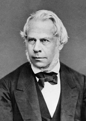
Many of the histological discoveries of Henle are described in the above. He classified tissues histologically. In the section on Gefässnerven (pp. 510, 690) Henle demonstrated the presence of smooth muscle in the end…
1842
#10381
Muséum d'anatomique pathologique de la Faculté de Médecine de Paris, ou Musée Dupuytren. Publié au nom de la Faculté. 2 vols. and atlas.
Plates lithographed after drawings by Émile Beau. Digital facsimile from BnFGallica at this link.
1844
#11148
Die männlichen und weiblichen Wollust-Organe des Menschen und einiger Säugetiere.
Kobelt provided the first comprehensive and accurate description of the function of the clitoris.Digital facsimile from digi.ub.uni-heidelberg at this link.
1845
#412
On a hitherto undescribed structure in the human hair sheath.
“Huxley’s layer” and “membrane” of the root sheath of hair follicles.
1845
#2294.1
Anatomical and pathological observations.
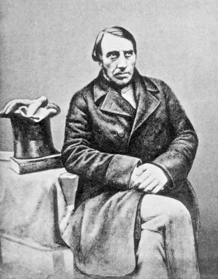
John Goodsir’s paper on “Centres of nutrition” anticipates to a certain extent the cell doctrine afterwards developed by Virchow (see No. 2299). Virchow dedicated the first edition of his Cellularpathologie to Goodsir…
1845
#13761
The origin of life: A popular treatise on the philosophy and physiology of reproduction, in plants and animals, including the details of human generation with a full description of the male and female organs. Illustrated by fine colored engravings on stone.
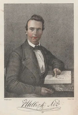
Digital facsimile of the 20th edition from Google Books at this link.
1846 –1849
#544
The microscopic anatomy of the human body, in health and disease.
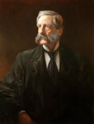
First English textbook on microscopical anatomy. His description of the concentric corpuscles of the thymus (p. 9) led to the term “Hassall’s corpuscles”.
1846
#413
Lehrbuch der Anatomie des Menschen.
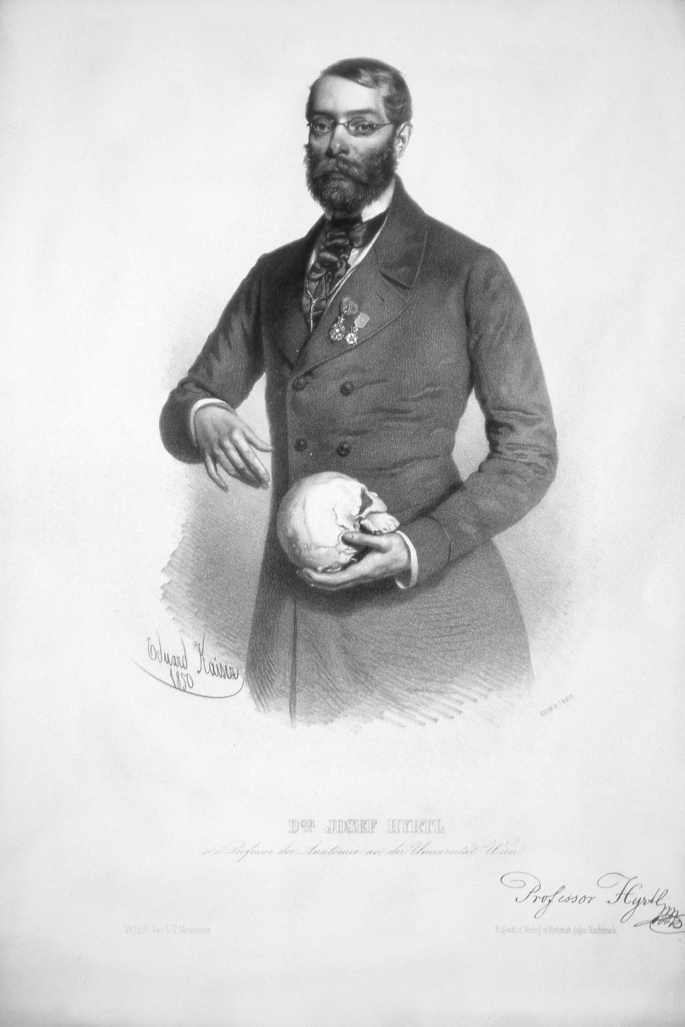
Hyrtl’s Lehrbuch passed through 22 editions and was translated into the principal modern languages.
1847
#994
Beobachtung einer tödlichen Peritonitis, als Folge einer Perforation des Wurmfortsatzes.
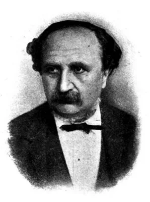
Description of “Gerlach’s valve”, sometimes seen at the orifice of the appendix.
1847
#414
Handbuch der topographischen Anatomie. 2 vols.
Hyrtl, professor of anatomy at Vienna, published the first text on topographical anatomy in German. He was for 30 years the most popular lecturer on the subject in Europe, and ranks as one of the greatest of medical s…
1848
#545
Bone or osseous tissue.
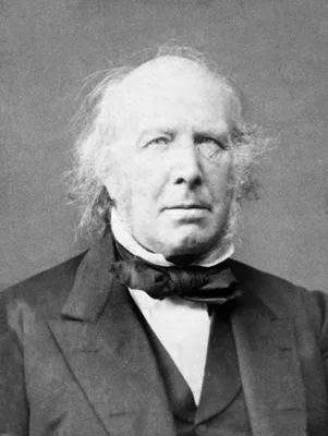
The discovery of the “fibers of Sharpey” is reported on pp. cxlii-cxliii.
1849
#1221
Ueber das Ligamentum pelvoprostaticum oder den Apparat, durch welchen die Harnblase, die Prostata und die Harnröhre an den untern Beckenöffnung befestigt sind.
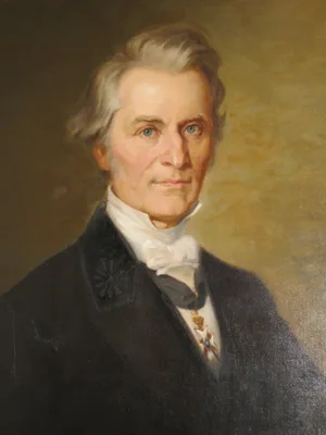
The “cave of Retzius” described.
1849
#13868
An atlas of anatomical plates of the human body, with descriptive letter-press in English and Hindustani.
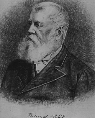
Digital facsimile from the Internet Archive at this link.
1850
#1222
Zur Anatomie der männlichen Geschlechtsorgane un Analdrüsen der Säugethiere.
Leydig was the first to describe the interstitial cells of the testis (“Leydig cells”).
1851
#13302
Surgical anatomy.
"The drawings of Maclise for Quain's Anatomy of the arteries and for his own Surgical anatomy are indeed done, as Quain wrote, with spirit and effect. These figures of anatomical dissection seem lifelike; in many plat…
1852
#415
A manual of artistic anatomy.
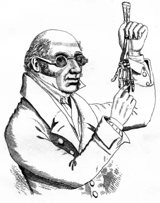
Knox, remembered because of his indiscreet association with the Edinburgh “resurrectionists”, was one of the best teachers of anatomy during the 19th century.
1852 –1859
#416
Anatome topographica sectionibus per corpus humanum congelatum triplici directione ductis illustrata. 8 pts.
Pirogov was the greatest of Russian surgeons. He introduced the teaching of applied topographical anatomy in Russia. His atlas of 220 plates represents the first use on a grand scale of frozen sections in anatomical i…
1853
#7241
Ueber einen neuen Muskel am Duodenum des Menschens, über elastische Sehnnen, und einige andere anatomische Verhãltnisse.
Treitz, a Czech pathologist, discovered the muscle at the duodenojejunal junction, later called “muscle of Treitz”; the fold of peritoneum over the muscle of Treitz is known as the "ligament of Treitz."
1854
#14061
Kitab fi tashrih beden al-insan [in Persian; English translation: Anatomy of the human body]. Lithographed text.
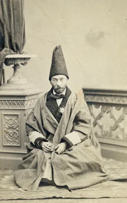
The first original Persian-language anatomy textbook based on western medical science, printed in a very small number of copies for the use of Polak’s Persian students. Polak, an Austrian physician, was responsible fo…
1855 –1871
#417
Handbuch der systematischen Anatomie des Menschen. 3 vols.
Considered by many authorities to be the greatest of the 19th-century systems of anatomy. Many structures are named after Henle, including the looped portion of the uriniferous tubules of the kidney, the layer of cell…
1855 –1869
#422
Medical anatomy: or, illustrations of the relative position and movements of the internal organs. 7 pts.
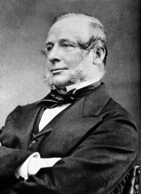
Sibson was professor of medicine at St. Mary’s Hospital. “Sibson’s fascia” and “muscle” are named after him. Plates 19-21 show movements, structure and sounds of the heart.
1857
#13248
Untersuchungen uber die Entwickelung des Schädelgrundes im gesunden und krankhaften Zustande und über Einfluss derselben auf Schädelform, Gesichtsbildung und Gehirnbau.
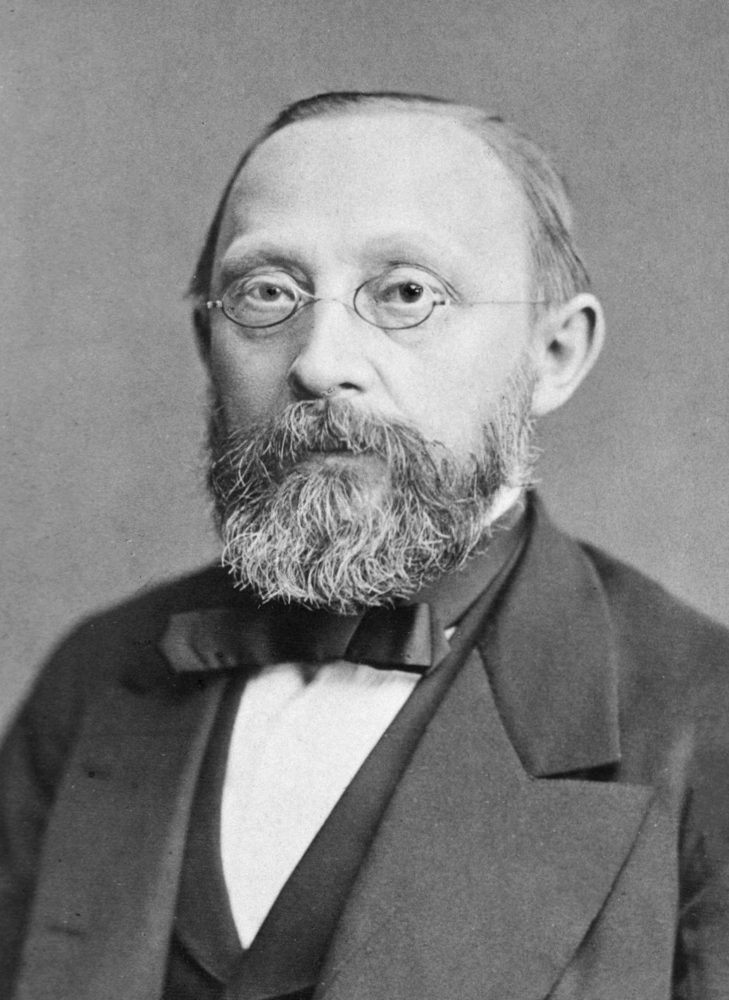
In his Investigations on the development of the base of the skull in healthy and diseased conditions, and on the influence of the same upon skull form, facial structure and brain formation Virchow laid the foundation …
1858
#418
Anatomy, descriptive and surgical
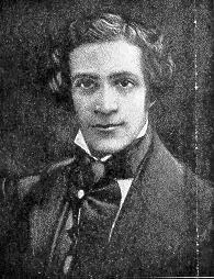
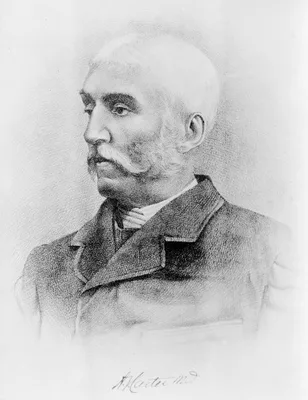
Gray’s textbook of anatomy remains today a standard work on the subject in the English-speaking world. The 37th edition appeared in 1989; the first American edition was published at Philadelphia, 1859. Digital facsimi…
1858
#419
A treatise on the human skeleton, including the joints.
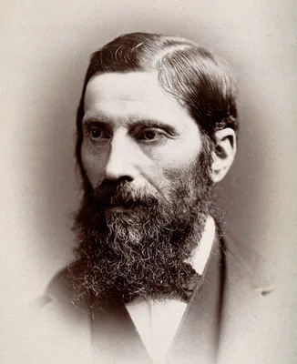
Humphry was professor of anatomy at Cambridge and became the first professor of surgery there. He founded the Journal of Anatomy and Physiology in 1867. “Humphry’s ligament” of the knee-joint is described on p. 546 of…
1858
#420
Des moyens chirurgicaux de favoriser la reproduction des os après les résections.
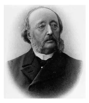
“Ollier’s layer”, the osteogenetic layer of the periosteum.
1861
#421
An elementary treatise on human anatomy.
Leidy illustrated this book himself. He was professor of anatomy at Philadelphia and the leading American anatomist of his time.
1861
#7608
Essays and observations on natural history, anatomy, physiology, psychology, and geology by John Hunter, F.R.S. Being his posthumous papers on those subjects, arranged and revised, with notes; to which are added the introductory lectures on the Hunterian collection of fossil remains delivered in the theatre of the Royal College of Surgeons of England, March 8th, 10th and 12th, 1855 by Richard Owen .... 2 vols.
Digital facsimiles from the Hathi Trust at this link.
1861 –1867
#14159
Atlas des peripherischen Nervensystems des menschlichen Körpers / Atlas du système nerveux périphérique du corps humain. Mit einem Vorwort von Prof. Dr. Th. W. L. Bischoff, nach der Natur photographirt von Joseph Alb. 10 parts.
Issued in 10 parts in folio (510 x 360 mm). Includes 46 large-format original photographs mounted on sheets of cardboard.
1862
#1108
Die Lymphgefässe und ihre Beziehung zum Bindegewebe.
“Recklinghausen’s canals”, the lymph canaliculi.
1862
#7621
Catalogue of the osteological portion of specimens contained in the Anatomical Museum of the University of Cambridge.
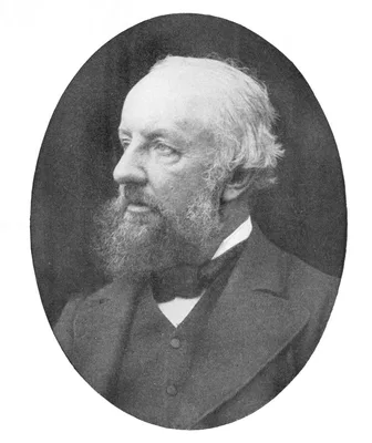
Digital facsimile from the Internet Archive at this link.
1864
#7331
The gray substance of the medulla oblongata and trapezium.
The first American medical book illustrated with photomechanically reproduced plates. Oliver Wendell Holmes praised the book for its remarkable photomicrographs, which may be the first published of brain cross-section…
1865
#10273
Wandtafeln zur Schwangerschafts- und Geburtskunde. Text volume in quarto format plus atlas in double elephant folio format (915 x 650mm.)
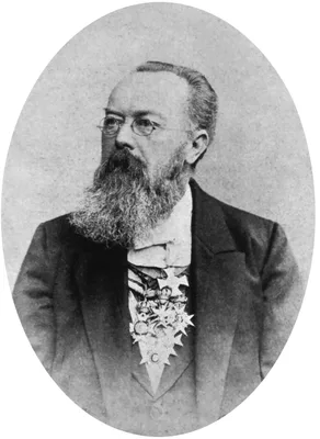
This huge atlas of obstetric wall charts contains 20 chromolithographed plates measuring over 3 feet by 2 feet, illustrating the female reproductive anatomy, stages of pregnancy, normal and breech presentations of the…
1868
#13221
The anatomical memoirs of John Goodsir F.R.S. Edited by William Turner. With a biographical memoir by Henry Lonsdale. 2 vols.
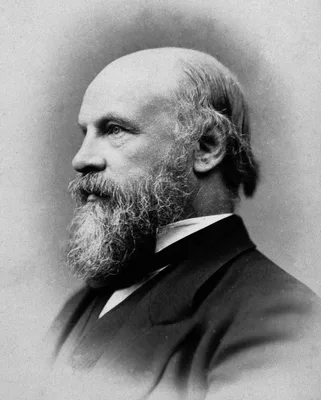
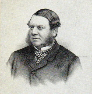
1869
#1009
Beiträge zur mikroskopischen Anatomie der Bauchspeicheldrüse. Inaugural-Dissertation.
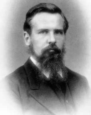
First account of the islets of Langerhans. In 1893 Édouard Laguesse attached the name of Langerhans to the structures. Langerhans did not suggest any function for them. The book was reprinted with an English translati…
1870
#423
Die descriptive und topographische Anatomie des Menschen.
1870
#491
Zur Kenntnis der menschlichen Placenta.
“Langhans’s layer”—the cytotrophoblast, the individual cells of which are termed “Langhans’s cells”.
1872
#424
Topographisch-anatomischer Atlas. Nach Durchschnitten angefrornen Cadavern.
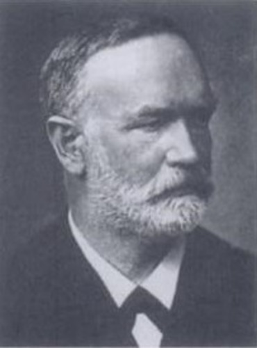
Fine illustrations of frozen sections. Translated into English by Edward Bellamy as An atlas of topographical anatomy after plane sections of frozen bodies. (Philadelphia: Lindsay and Blakiston, 1877).
1872
#6188
Die Lage des Uterus und Foetus am Ende der Schwangerschaft nach Durchschnitten an gefrornen Cadavern
Supplement to No. 424.
1873
#425
Die Corrosions-Anatomie und ihre Ergebnisse: mit 18 chromolithographirten Tafeln.
Hyrtl significantly enhanced the techniques of corrosion anatomy, a technique of preparing anatomical specimens invented by Frederik Ruysch. He built up a collection unsurpassed in Europe. In this work Hyrtl described…
1876
#552
Ueber Stemzellen der Leber.
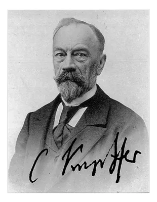
“Kupffer cells” – macrophage stellate cells in the lining of the blood channels in the liver. Confocal microscopy picture showing the steady-state location and interactions between Kupffer cells (Red), hepatic stellat…
1876
#2304
Die Sections-Technik im Leichenhause des Charité-Krankenhauses.
On the technique of dissection. English translation, London, 1876.
1878
#1012.1
Ueber die Pepsinbildung in den Pylorusdrusen.
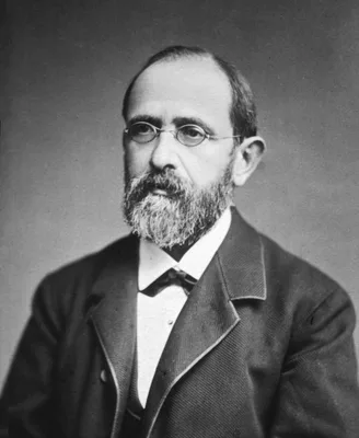
Heidenhain pouch.
1881
#7645
Frozen sections of a child by Thomas Dwight. Fifteen drawings from nature by H. P. Quincy.
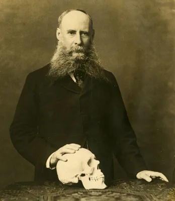
The first atlas of cross-sectional anatomy published in the United States. Digital facsimile from the Internet Archive at this link