
Facets
Browse across eight MeSH (opens in new tab) facets — era, geography, science, specialty, technology, history, culture, and reference. Select one tag per group; counts update across the others. What’s new in facet browse how facets relate to subjects and MeSH.
Clear filtersFacet filters
Geography
Specialties & Disease
- Anatomy & Pathology 59
- Cardiology & Blood 48
- Neurology & Psychiatry 49
- Obstetrics & Reproductive 10
- Infectious Disease (General) 2
- Surgery & Anesthesia 14
- Public Health 31
- Immunology & Dermatology 60
- General Clinical Medicine 169
- Military Medicine 8
- Psychology 9
- Alternative & Fringe Medicine 3
- Pediatrics 3
- Ophthalmology & Vision 26
- ENT & Hearing 19
- Urology & Nephrology 13
- Gastroenterology & Hepatology 10
- Pulmonary & Respiratory 5
- Rheumatology, Rehab & Pain 3
- Internal, Emergency & Geriatric 1
- Veterinary Medicine 1
- Epidemiology & Demography 31
- Physiology & Embryology 34
- Dentistry 8
- Plagues & Epidemics 55
- Microbiology & Virology 49
Social & Historical Studies
Institutions & Culture
Reference & Scholarly Works
13 entries match Urology & Nephrology [C13] · Diagnostics & Imaging [E01]
1894
#8970
Kystophotographischer Atlas.
Digital facsimile from the Internet Archive at this link.
1896
#3636
Calculs du rein. Calculs de la vésicule biliaire.
Chappuis and Chauvel were the first to study biliary concretions by means of Roentgen rays.
1896
#4293
Roentgen rays. Photography of renal calculus.
First radiogram of renal calculus.
1899
#4191.1
Sonde urétérale opaque. In Traité de chirurgie, 2e ed. vol. 7, p. 414.
Tuffier made the first pioneer attempt to visualize the urinary tract by the combination of an opaque ureteral styletted catheter and radiography.
1901
#4294
Uretercatheterezés és radiographia.
Géza von Illyés showed that ureteral calculi could be accurately demonstrated by x rays with the help of an indwelling opaque catheter. German translation in Dtsch. Z. Chir., 1901, 62, 132-40.
1903
#4191.3
Zur Technik der Röntgenphotographie (Lendenwirbel, Blasensteine).
Cystography. Wittek filled the bladder with air and was able to demonstrate vertebrae and urinary calculus.
1905
#4192
Die Gestalt der menschlichen Harnblase im Röntgenbilde.
First cystograms.
1906
#4231
Pyelographie (Roentgenographie des Nierenbeckens nach Kollargolfüllung).
Introduction of pyelography.
1920
#4198
X-ray studies of the seminal vesicles and vasa deferentia after urethroscopic injection of the ejaculatory ducts with thorium – a new diagnostic method.
Vesiculography first demonstrated.
1921
#4241
Un nuevo procedimiento para explorar al riñón.
Perirenal insufflation of oxygen, for the roentgenological study of the kidney.
1923
#4199
Roentgenography of the urinary tract during excretion of sodium iodide.
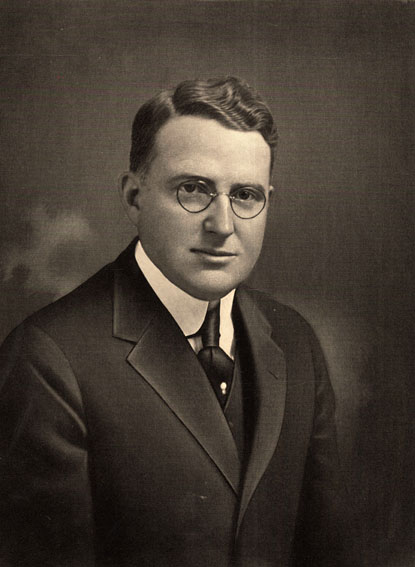
First use of sodium iodide in uretero-pyelography. With C. G. Sutherland and A. J. Scholl.
1928 –1929
#4246
Studies of urea excretion.
Blood urea clearance test. With J. F. Mcintosh and D. D. Van Slyke.
1954
#4256.2
Nephrotomography. A preliminary report.
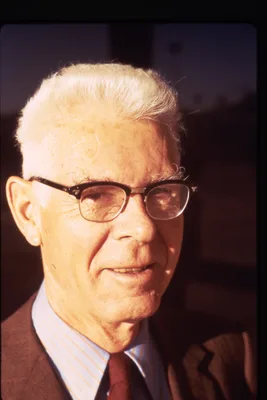
With W. Dubilier and J. C. Monteith.